|
From Wikipedia the free encyclopedia, by MultiMedia |
| Sacrospinalis muscle |
|---|
|
|
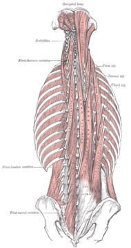
| Deep muscles of the back. |
Sacrospinalis is a very thick, lateral portion of an epaxial muscle in mammals which continues anteriorly up to the neck and divides into three muscles: semispinalis, longissimus, and iliocostalis. Its origin is on the spines of the last four thoracic vertebrae, and its insertion is on both the spines of the most craniad thoracic vertebrae and the cervical vertebrae. Its action is to extend the vertebral column.
The semispinalis is the muscle immediately lateral to the multifidus spinae and is the most medial of all three. It consists of diagonal fibers.
The longissimus is the muscle lateral to the semispinalis. It is the longest subdivision of the sacrospinalis that extends forward into the transverse processes of the posterior cervical vertebrae.
The iliocostalis is the muscle immediately lateral to the longissimus that is the nearest to the furrow that separates the epaxial msucles from the hypaxial. It lies very deep to the fleshy portion of the serratus ventralis.
The Sacrospinalis (Erector spinæ), and its prolongations in the thoracic and cervical regions, lie in the groove on the side of the vertebral column. They are covered in the lumbar and thoracic regions by the lumbodorsal fascia, and in the cervical region by the nuchal fascia. This large muscular and tendinous mass varies in size and structure at different parts of the vertebral column. In the sacral region it is narrow and pointed, and at its origin chiefly tendinous in structure. In the lumbar region it is larger, and forms a thick fleshy mass which, on being followed upward, is subdivided into three columns; these gradually diminish in size as they ascend to be inserted into the vertebræ and ribs.
The Sacrospinalis arises from the anterior surface of a broad and thick tendon, which is attached to the medial crest of the sacrum, to the spinous processes of the lumbar and the eleventh and twelfth thoracic vertebræ, and the supraspinal ligament, to the back part of the inner lip of the iliac crests and to the lateral crests of the sacrum, where it blends with the sacrotuberous and posterior sacroiliac ligaments. Some of its fibers are continuous with the fibers of origin of the Glutæus maximus. The muscular fibers form a large fleshy mass which splits, in the upper lumbar region into three columns, viz., a lateral, the Iliocostalis, an intermediate, the Longissimus, and a medial, the Spinalis. Each of these consists from below upward, of three parts, as follows:
| Lateral Column | Intermediate Column | Medial Column |
| Iliocostalis | Longissimus | Spinalis |
| I. lumborum | L. dorsi | S. dorsi |
| I. dorsi | L. cervicis | S. cervicis |
| I. cervicis | L. capitis | S. capitis |
The Iliocostalis lumborum (Iliocostalis muscle; Sacrolumbalis muscle) is inserted, by six or seven flattened tendons, into the inferior borders of the angles of the lower six or seven ribs.
The Iliocostalis dorsi (Musculus accessorius) arises by flattened tendons from the upper borders of the angles of the lower six ribs medial to the tendons of insertion of the Iliocostalis lumborum; these become muscular, and are inserted into the upper borders of the angles of the upper six ribs and into the back of the transverse process of the seventh cervical vertebra.
The Iliocostalis cervicis (Cervicalis ascendens) arises from the angles of the third, fourth, fifth, and sixth ribs, and is inserted into the posterior tubercles of the transverse processes of the fourth, fifth, and sixth cervical vertebræ.
The Longissimus dorsi is the intermediate and largest of the continuations of the Sacrospinalis. In the lumbar region, where it is as yet blended with the Iliocostalis lumborum, some of its fibers are attached to the whole length of the posterior surfaces of the transverse processes and the accessory processes of the lumbar vertebræ, and to the anterior layer of the lumbodorsal fascia. In the thoracic region it is inserted, by rounded tendons, into the tips of the transverse processes of all the thoracic vertebræ, and by fleshy processes into the lower nine or ten ribs between their tubercles and angles.
The Longissimus cervicis (Transversalis cervicis), situated medial to the Longissimus dorsi, arises by long thin tendons from the summits of the transverse processes of the upper four or five thoracic vertebræ, and is inserted by similar tendons into the posterior tubercles of the transverse processes of the cervical vertebræ from the second to the sixth inclusive.
The Longissimus capitis (Trachelomastoid muscle) lies medial to the Longissimus cervicis, between it and the Semispinalis capitis. It arises by tendons from the transverse processes of the upper four or five thoracic vertebræ, and the articular processes of the lower three or four cervical vertebræ, and is inserted into the posterior margin of the mastoid process, beneath the Splenius capitis and Sternocleidomastoideus. It is almost always crossed by a tendinous intersection near its insertion.
The Spinalis dorsi, the medial continuation of the Sacrospinalis, is scarcely separable as a distinct muscle. It is situated at the medial side of the Longissimusdorsi, and is intimately blended with it; it arises by three or four tendons from the spinous processes of the first two lumbar and the last two thoracic vertebræ: these, uniting, form a small muscle which is inserted by separate tendons into the spinous processes of the upper thoracic vertebræ, the number varying from four to eight. It is intimately united with the Semispinalis dorsi, situated beneath it.
The Spinalis cervicis (Spinalis colli) is an inconstant muscle, which arises from the lower part of the ligamentum nuchæ, the spinous process of the seventh cervical, and sometimes from the spinous processes of the first and second thoracic vertebræ, and is inserted into the spinous process of the axis, and occasionally into the spinous processes of the two vertebræ below it.
The Spinalis capitis (Biventer cervicis) is usually inseparably connected with the Semispinalis capitis.
Cats, made by MultiMedia | Free content and software
This guide is licensed under the GNU Free Documentation License. It uses material from the Wikipedia.